

The Biomedical Imaging Center (BIC) is an interdisciplinary, multimodal facility supporting human and preclinical research across three UT Austin sites. Researchers have access to MRI/MRS, PET, CT, SPECT, and optical imaging systems, with options for independent or assisted use, training, and centralized data archiving and analysis support.
Quick Links
Getting Started
Step 1: Consultation for New Users and New Projects
All new users and current users/collaborators beginning a new project must start with a consultation. Please contact the Facility Director to arrange an initial discussion of your research project or program.
This consultation helps determine feasibility, requirements, approvals, and next steps before access is granted.
A new project includes any research study that involves:
- A new or updated funding source (including grant extensions)
- A new or updated IRB or IACUC protocol (excluding personnel-only updates)
- Development of a new scan protocol, or studies involving more than 10 sessions
Step 2: Request Access to the Facility Billing System (FBS)
BIC uses the FBS to schedule services and instrument use.
Once approved through the consultation process, new users may request access to FBS by submitting the FBS Access Request Form. Access to FBS is required before scheduling any services or instrumentation.
Step 3: Request Training or Technical Support
Existing users and collaborators may request training, technical assistance, or operational support by submitting a BIC Training and Support Request Form.
This includes requests related to instrument training, troubleshooting, or assistance with approved and active projects.
Acknowledgment of Core Services and Equipment
Please support the mission of the BIC by acknowledging the use of our facility, equipment and resources using our unique research resource identifier number (RRID:SCR_021898). Citation by you will ensure the BIC is recognized as a key resource at UT Austin and will promote institutional support and future funding.
RRID:SCR_021898
Locations
BIC operates three imaging facilities on the UT Austin campus:
Imaging Research Center (NHB)
The Imaging Research Center (IRC) is the original site of the BIC located in the Norman Hackerman Building (NHB) in the middle of main campus. The IRC is a ~6,000 square foot facility dedicated to state-of-the-art neuroimaging and features BIC’s flagship Siemens 3T Prisma MRI scanner (installed Summer 2023). The site was renovated following the installation of the Prisma and includes the infrastructure required to support human MRI studies, including:
- two interview rooms
- exam/study prep room
- BSL-2 lab space
- participant changing room
- communal workspace/conference room
- electronics workshop
- wet-lab space
- prep-room for preclinical imaging
BIC staff offices are located within the footprint of the IRC and parking spaces for human research participants are located nearby on Speedway.
UT Austin Imaging Center (HDB)
The UT Austin Imaging Center is a ~15,000 sq. ft. clinical / research facility located in the Health Discovery Building (HDB) at the Dell Medical School campus. The UTAIC supports human imaging research, out-patient diagnostic imaging (delivered by our partners at UT Health Austin Advanced Imaging), and facilitates clinical-research studies. The site hosts a Siemens 3T Vida MRI scanner and a GE Discovery MI PET/CT scanner and features all the standard infrastructure to support human imaging research, including:
- three interview / study rooms; BSL-2 lab space
- four changing rooms
- four patient prep rooms
- electronics workshop
- communal workspace
- prep room for preclinical imaging
BIC staff offices are on-site; participant parking spaces for research imaging studies are in the adjacent Health Center Garage (HCG).
Preclinical Imaging Suite (HDB)
The Preclinical Imaging Suite (PIS) is a ~2,000 sq. ft. facility dedicated to preclinical imaging located within the vivarium in the Health Discovery Building (HDB). The site houses preclinical imaging systems for MRI, SPECT/CT/PET, and optical imaging as well as an X-ray irradiator. Full resources for preclinical imaging are provided including adjacent procedure rooms for study preparation and each imaging system is supported by the necessary peripheral equipment for in-vivo studies.
Satellite Sites (SEA and ARC)
Additional resources are available at satellite sites in the Seay Building (SEA) and the Animal Resource Center (ARC):
- Seay Building: hosts an MRI Simulator mock scanner adjacent to our collaborators in the Department of Psychology to facilitate participant preparation and acclimation.
- Animal Resource Center: hosts a MultiRad 225 X-ray irradiator to complement the system located in the Preclinical Imaging Suite.
Services
BIC supports both independent and assisted use of its imaging systems and provides the following services:
- Initial consultation on establishing an imaging research project or program at BIC
- Development / implementation of MRI / fMRI protocols on our 3T and 7T systems
- Ongoing project advice and technical support for the imaging systems and ancillary equipment
- Educational courses on imaging physics and technology
- Training in MRI scanner safety (MRI Level 1 certification) and scanner-operation (MRI Level 2 certification)
- Training in scanner operation for our SPECT/CT/PET and optical imaging systems and the use of our X-ray irradiators
- BIC staff may be available are available to assist with human / preclinical imaging studies
- MRI and PET/CT Technologists are available to operate the Siemens Vida and GE Discovery systems in HDB
- Assistance with IRB and IACUC protocol development
- Post-study support for data archiving, management and common analyses
Instrumentation
Human Imaging Scanners
Siemens Prisma 3.0T MRI (located at the Imaging Research Center in NHB)
The Prisma is BIC’s flagship 3T MRI system and is configured for state-of-the-art neuroimaging research. It features a highly homogeneous magnet as well as high-performance XA line electronics and software, and the XR gradient set offers a maximum amplitude of 80mT/m across all axes with a slew-rate of 200mT/m/s. The system is equipped with a high-power 2nd order shim-set, multi-nuclear capabilities, a full complement of RF coils for brain, body and extremity imaging, and an extensive library of standard and advanced pulse sequences. Investigator- and 3rd party-developed sequences may also be installed under BIC’s Master Research Agreement with Siemens, as well as manufacturer Works-in-Progress (WIP) protocols.
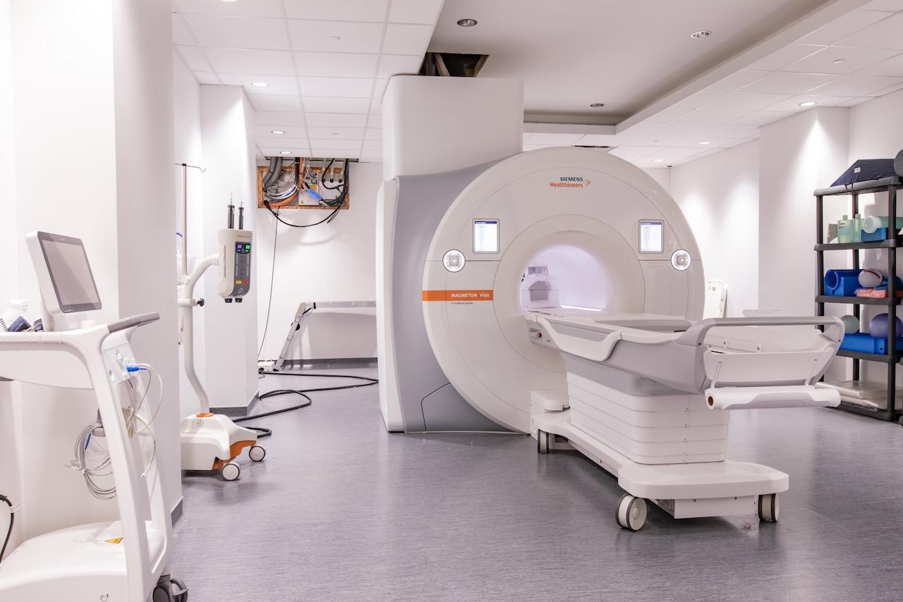
Siemens Vida 3.0T MRI (located at the UT Austin Imaging Center in HDB)
The Vida is a wide-bore 3T MRI scanner configured for clinical and research imaging. It is capable of high-resolution anatomical and functional MRI and is equipped with a broad array of RF coils for brain, body and extremity imaging. The system also features XA line electronics and software including BioMatrix technology for integrated physiological triggering without peripheral devices. An extensive library of standard and advanced pulse sequences is available and the system is also covered by BIC’s Master Research Agreement enabling the installation of investigator-developed and 3rd party pulse sequences.
GE Discovery MI PET-CT (located at the UT Austin Imaging Center in HDB)
The GE Discovery PET-CT scanner is capable of high-resolution, research and clinical imaging. In CT mode, the system has a 50cm diagnostic FOV which can expand to 70cm for attenuation correction and is capable of ultra-low pitch settings for large axial coverage with minimal gantry rotations. Available scan modes include standard Axial, helical, and cine scan modes are available along with iterative image reconstruction techniques enabling high resolution with low X-ray doses. The PET subsystem features lutetium-based detectors integrated to a fully digital acquisition enabling high sensitivity + ultra-low noise image reconstruction plus time-of-flight detection capabilities.
Mock MRI Scanners
- An MRI Simulator PST-101728 is located in Seay Building adjacent to our collaborators in the Department of Psychology. This system supports participant acclimation to the MRI environment and features a 60cm bore with tapered opening (comparable to our Siemens Prisma scanner), a movable patient table and realistic mock head-coil, and the capability to mimic gradient noise and fMRI audio and visual stimuli.
- A mock GE Signa will soon be installed within BIC’s facility at the Health Discovery Building. This system is the shell of BIC’s original 1.5T MRI scanner and features the original 70 cm bore, patient table and a subset of RF coils.
Peripheral Equipment
- fMRI Response Devices: Both the Siemens Prisma and Vida MRI scanners are equipped with Current Designs packages to record participant responses during fMRI studies. Each system includes a FIU-932 interface supplemented with a range of fiber-optically linked single and bi-manual button-box response devices. The 932 interface accepts the TTL trigger output from the scanner for experimental synchronization and triggering and outputs response data via USB.
- Video Systems: Visual stimulation for fMRI studies is provided by NordicNeuroLab products. The Siemens Prisma and Vida MRI scanners are each equipped with the MR-compatible InroomViewingDevice, a 40 inch TFT LCD display that supports both ultra-high (4k, UHD) and standard HD (1080p) resolution. A VisualSystemHD package is also available for advanced applications requiring higher-quality graphics. MediGlasses kits are also provided for MR-safe prescription glasses requirements.
- Audio Systems: In addition to the built-in headphone systems of the human imaging systems, the Siemens MRI scanners are equipped with high-quality systems for auditory stimulation. Both the Prisma and Vida have recently been upgraded to the Optoacoustics OptoACTIVE III package which provides mechanical isolation and active noise cancellation for the headphones and microphone subsystems, permitting high-fidelity audio and the recording of spoken responses while scanning. Sensimetrics S15 earphone systems are also available.
- Physiological Monitoring / Intervention: Both the Prisma and Vida scanners are equipped with Siemens OEM accessories for physiological monitoring of ECG, respiration and SpO2, with integrated logging of physiological data. Additional physiological monitoring / intervention capabilities are provided with multimodal BioPac MP200 (Prisma) and MP160 (Vida) systems, enabling MRI compatible measurements of ECG, respiration, skin-conductance and electrical stimulation with the ability to expand to other modalities as required.
- Contrast Injections: Power injectors for the delivery of contrast agents / tracers* are available on each human imaging system.
*Appropriate medical coverage required for the provision of any exogenous agent.
Preclinical Imaging Scanners
Bruker BioSpec 7.0T MRI (located at the Preclinical Imaging Suite in HDB)
Preclinical MRI at BIC is supported by our 7T Bruker BioSpec scanner. This system has a high duty-cycle gradient coil w/ 9cm free-bore; higher-order shimset and both 1H and X-nucleus capabilities. 1H volume coils are available in a variety of diameters to enable phantom, organ and whole-body imaging, as well as a volume-transmit / surface-receive coil setup for high-sensitivity applications. Our collaborators in the Que group also have a variety of coils for 19F applications. The system is equipped with peripheral equipment for anesthesia delivery, physiological monitoring, infusions and other interventions.
Siemens Inveon PET/CT (located at the Preclinical Imaging Suite in HDB)
PET and CT imaging is supported by a Siemens Inveon MM scanner. For CT mode, the system is equipped with a variable-focus X-ray source and 165mm detector enabling resolution down to ~7um isotropic. Moving table and iterative reconstruction increase the axial field-of-view to enable whole animal imaging. In PET modem the system has 4 x 16 rings of detectors (20 x 20 LSO arrays) offering resolution to 1.6 x 1.6mm. Anesthesia and full physiological monitoring capabilities are also available. This system is scheduled to be upgraded to a Mediso NanoScan SPECT/CT/PET system during 2026, a detailed scanner configuration coming soon
Mediso NanoScan SPECT/CT/PET (located at the Preclinical Imaging Suite in HDB)
Content to be added
Xenogen IVIS Spectrum (located at the Preclinical Imaging Suite in HDB)
The IVIS is an optical imaging system capable of imaging bioluminescent / fluorescent reporters. Multiple excitation and emission filters are available enabling detection of blue to near-IR wavelengths. The system is capable of spectral unmixing so multiple fluorescent probes within same sample can be detected and separated. The system was upgraded during 2025 and features a new optical camera / chiller assembly, electronics boards and host computer. Isoflurane anesthesia is integrated into the system, and a multi-channel manifold permits simultaneous imaging of up to 5 specimens.
MultiRad 350 X-ray Irradiator (located at the Preclinical Imaging Suite in HDB)
The MultiRad350 is a compact, fully-shielded, self-contained x-ray irradiator designed for high-dose and uniform depth irradiation, making it ideal for dense biological specimens and a broad range of industrial applications. It features an adjustable sample shelf, software-controlled turntable and an integrated dosimeter with Automatic Dose Control (ADC). A touchscreen interface with easy-to-use software controls all parameters and enables programming of doses from 10 – 350 kVp.
MultiRad 255 X-ray Irradiator (located at the Animal Resources Center)
The MultiRad225 is mid-powered x-ray irradiator suitable for targeted irradiation of small samples. Like its more powerful sibling, it is a fully-shielded, self-contained unit an adjustable sample shelf, software-controlled turntable and an integrated dosimeter with Automatic Dose Control (ADC). Programming of doses from 10 – 225 kVp is possible via the touchscreen interface and easy-to-use software.
Data Archiving and Analysis
PACS Archive: Research imaging data from all primary modalities (MRI / SPECT / CT / PET) is archived to our communal data server. BIC_PACS offers ~200TB of expandable image storage and data is directly allocated into user-specific lab directories which can be SAMBA mounted. This service is incorporated into BIC’s instrumentation fees and is available to all users free of charge. Advanced archiving pipelines are also available.
Compute Server: BIC’s image acquisition and archiving capabilities are augmented by our compute server, which is available via an annual user-fee per login. bicfcomp features dual 28-core / 56-thread CPU’s with 768GB RAM and a 2TB ultra-high-speed SSD, fiber-optically linked to BIC_PACS for ultra-fast I/O. It supports common image analysis pipelines (including GUI capabilities) via singularity and docker – more powerful than a lab or department-based server, more user-friendly than UT’s advanced computing capabilities at the TACC.
Community
BIC supports the research programs of over 80 independent investigator-led groups spread across 23 Academic Departments and eight Colleges and Schools. Areas of interest include:
- the neural anatomy, physiology, and cognitive function associated with memory, aging, speech and language, learning and development, and neurological disorders such as Alzheimer’s disease and MS
- the pathophysiology of TBI and other brain injuries
- the neural mechanisms underpinning bipolar disorder, PTSD, fear, addiction, and other psychiatric disorders
- characterization of tumor growth and treatment response in breast, brain and prostate cancer using advanced quantitative-MRI (qMRI) methods
- the genetic determinants of early brain development and the impacts of cancer and chemotherapy-related neurodegeneration on cognition
- abdominal MRI to assess maternal / infant nutrition and establish biomarkers for diabetes and pancreatic cancer
- the development of novel MRI methods to enhance the efficiency of image acquisition and the measurement of blood flow and oximetry
- the creation of novel contrast mechanisms using 19F-labelled nano-particles
- the regulation and function of stem cells, tissue engineering to treat injuries to the nervous system, and the development of neuroprosthetics
- ongoing clinical trials in partnership with the Departments of Oncology and Neurology at the Dell Medical School
Additional Resources
Education and Training
BIC supports a range of undergraduate and graduate level courses at UT Austin, and offers its own courses in imaging physics, scanner safety and scanner operation for affiliated researchers. BIC also provides didactic support for the Dell Medical School Radiology Residency program and is a host site for the UT Austin in Portugal program.
Research Participants
If you are enrolled in an ongoing research study, please contact the Principal Investigator or study coordinator directly. Inquiries about prospective involvement in a research study should also be directed to the research team performing the study.
Research Compliance
Support for the development of IRB or IACUC protocols for imaging research projects is also available, including standardized language to ensure compliance and conformation with BIC and institutional policies and guidelines.
Staff
Douglas Befroy, Ph.D. - Director
Doug Befroy is the Director of the Biomedical Imaging Center. He has more than 20 years of experience developing advanced MR methods to investigate physiology and metabolism in human and animal models. He received his Ph.D. at Oxford University after which he joined the imaging community in the Departments of Diagnostic Radiology and Internal Medicine at Yale University School of Medicine. At Yale, Befroy served in several leadership roles, including acting as Director of the Core of Muscle and Liver Spectroscopy and Co-Director of the Mouse Imaging Core. Most recently, he founded and ran an independent consultancy firm that provides technical support and innovative research solutions to imaging centers and scientists across the globe.
Cici Cumba - Senior Software Engineer
Email: cici.cumba@austin.utexas.edu