Systems
Human Imaging Scanners
-
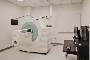
Siemens Skyra 3.0T MRI (located in NHB)
Show More
-
The Siemens Skyra 3T enables high-resolution imaging capabilities at the UT bic. This whole-body 3.0T device is configured with 64 receiver channels. It is capable of integrated parallel acquisition techniques and provides higher signal to noise in the parallel imaging mode than its predecessors. The maximum acceleration factor using parallel imaging is 16 using either mSENSE or GRAPPA, and 3D scanning can be accelerated in two directions (maximum acceleration factor of 4 in second direction). The gradients of the Skyra have a maximum amplitude of 45 mT/m and a maximum slew rate of 200 T×m-1×s-1, yielding a minimum rise time of 225μs. The vector gradient performance (vector summation of all three gradient axes) results in a maximum effective amplitude of 78 mT/m and a maximal effective slew rate of 346 T/m/s. All three gradient coils are force-compensated to reduce vibration and deliver superior eddy current performance. The water-cooled gradient amplifier has a maximum amplitude potential of 2,250 volts and a maximum current output of 750 amps. The instrument has a minimum slice thickness (in two dimensions) of 0.1 mm and a minimum partition thickness (in three dimensions) of 0.05 mm. The instrument produces high sensitivity, with main field, or B0, homogeneity of 1.4 ppm VRMS for a 40 cm diameter spherical volume. Single shot EPI sequences for measuring diffusion-weighted data sets with up to 256 directions of diffusion weighting are also a part of this instrument’s capability. It provides diffusion tensor imaging and parametric maps derived from fractional anisotropy calculated in real time, automatically. Additional sequence options include Arterial Spin Labeling, and susceptibility weighted imaging (SWI) with both fully supported with parametric and phase map reconstructions.
Response Devices
The bic uses a 932 Package provided by Current Designs, Philadelphia, PA. Currently, there is a four button bimanual box (2×2) available and an eight button bimanual box (2×4). The 932 interface accepts the TTL trigger output from the Skyra during EPI experiments for experiment and response synchronization.Visual Stimulation
The PROPixx projector at the bic has a native resolution of 1920 x 1080 and can be reliably driven with refresh rates up to 500 Hz. It uses high brightness LEDs as a light source, giving a larger color gamut and much longer lifetime than conventional bulbs. For stereo vision applications, a high speed circular polarizer allows for 3D displays using passive polarizing glasses, with interleaved binocular refresh rates up to 400 Hz.The PROPixx also synchronizes virtually any input or output devices to the timings of the video signal with microsecond precision. These can include stereo audio, button boxes for precise reaction-time measurement, triggers for electrophysiology and eye-tracking equipment, and/or any custom analog I/O subsystem. You can now successfully synchronize all of your subject I/O to video refresh with microsecond precision. It is field upgradeable, allowing for custom features to be added via firmware update.
A software toolbox, PLDAPS, is shared by UT researchers and can be used in MATLAB (with the Psychophysics Toolbox) to take advantage of these functionalities with minimal additional programming overhead.
Physiological Monitoring and Recording
The Skyra is equipped with its own accessories for physiological monitoring and triggering. Users can select a 2, 3, or 4-lead ECG configuration. The acquired cardiac waveforms can be used to trigger most sequences or they can be recorded for retrospective image reconstruction and/or analysis. For studies not needing to trigger to a precise phase in the cardiac cycle, a plethysmographic finger clip can be used instead. This can provide adequate triggering for cine cardiac imaging without the need to attach ECG leads which some subjects see as overly intrusive. This method is also capable of providing real-time pulse rate and retrospective analysis. Similar triggering and recording options are available via respiratory bellows that are strapped to the subject’s abdomen over their clothing.Auditory Systems
In addition to the built-in headphone system of the MRI scanner, an Optoacoustics OptoACTIVE system is available, which provides active noise cancellation for both headphones and microphone, allowing high-fidelity stimulus presentation as well as a collection of spoken responses.Liquid Delivery
A gustometer system (designed by Dr. Eric Stice) is available for studies involving liquid delivery. The gustometer system is a portable device that consists of a laptop computer that can control up 7 independently programmable BS-8000 syringe pumps to deliver precise amounts of liquid stimulus to the supine subject at precisely timed intervals and durations. The pumps, which infuse liquids at rates of 6- 15 mL/min, are controlled by programs written using MATLAB. The gustometer mouthpiece is a collar capable of holding up to 7 beverage tubes and was designed to deliver tastants or tasteless solution to a consistent place on the tongue. Each tube is attached to one-way syringe activated check valves to prevent dripping and participants sucking on the tubes. -
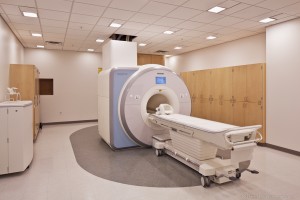
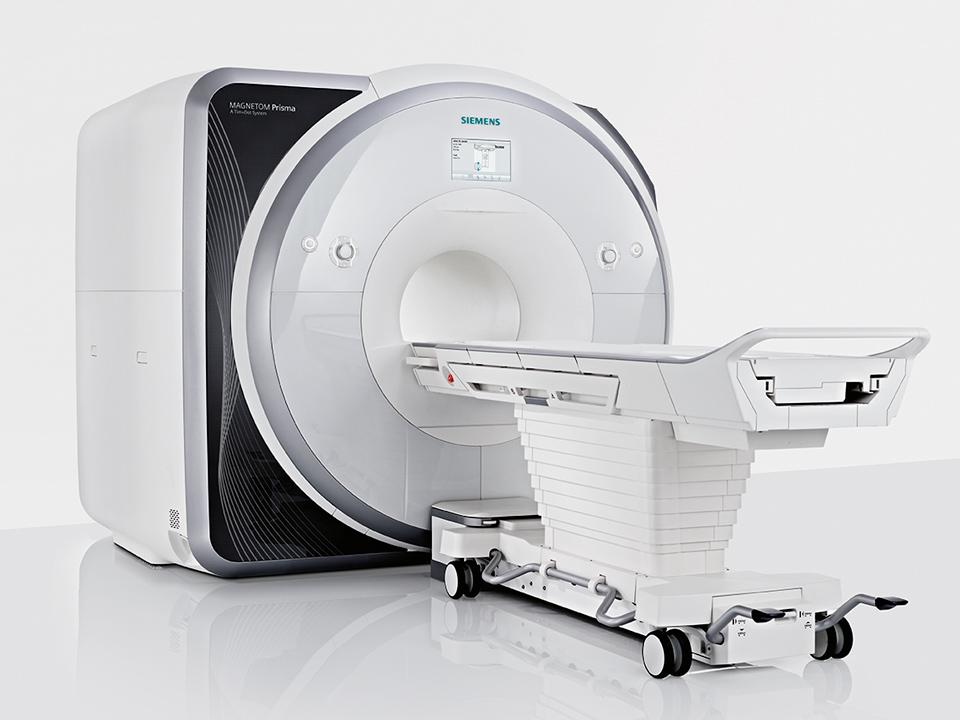
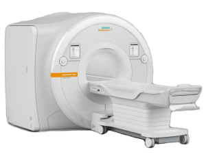
Siemens Prisma 3.0T MRI (to be located in NHB)
Show More
-
BIC will be upgrading our 3T Skyra system in NHB to a 3T Prisma in March 2023. The Prisma is Siemens’ state-of-the-art 3T system for neuroimaging research. It features a highly homogeneous magnet, the XR gradient set offering a maximum amplitude of 80mT/m across all axes with a slew-rate of 200mT/m/s and high-performance XA line electronics and software. The system will be equipped with a high-power 2nd order shim-set, multi-nuclear capabilities, a full complement of RF coils for brain, body and extremity imaging, and an extensive library of standard and advanced pulse sequences. The Prisma will provide a significant performance upgrade on our Skyra (and Vida) systems with improved signal/noise – especially in hard to shim regions – and enhanced gradient performance leading to improved image quality and/or higher spatial / temporal resolution. Integrating this scanner into BIC’s fleet of imaging systems will allow the Center to match our users’ needs for the next 10+ years and enable our collaborators to maintain and extend their innovative MRI / fMRI research programs.
-
Siemens Vida 3.0T MRI (located in HDB)
Show More
-
The Siemens Vida 3T enables high-resolution imaging capabilities at the UT bic. This whole-body 3.0T device is configured with 64 receiver channels. It is capable of integrated parallel acquisition techniques and provides higher signal to noise in the parallel imaging mode than its predecessors. The maximum acceleration factor using parallel imaging is 16 using either mSENSE or GRAPPA, and 3D scanning can be accelerated in two directions (maximum acceleration factor of 4 in the second direction). The gradients of the Vida have a maximum amplitude of 60 mT/m and a maximum slew rate of 200 T×m-1×s-1, yielding a minimum rise time of 300μs. All three gradient coils are force-compensated to reduce vibration and deliver superior eddy current performance. The water-cooled gradient amplifiers have a maximum potential of 2.7 megawatts that can be distributed across any of the 3 axes. Single shot EPI sequences for measuring diffusion-weighted data sets with up to 512 directions of diffusion weighting are also a part of this instrument’s capability. It provides diffusion tensor imaging and parametric maps derived from fractional anisotropy calculated in real time, automatically. Additional sequence options include 2D and 3D Arterial Spin Labeling, LiverLab, and susceptibility weighted imaging (SWI) with both fully supported with parametric and phase map reconstructions. Compressed sensing is available for some free-breathing techniques. The Vida makes use of a feature Siemens calls BioMatrix that allows for, among other things, respiratory triggering without the use of a bellows as well as an extra shim coil built into both the 20 and 64 channel head coils.
Response Devices
The bic uses a 932 Package provided by Current Designs, Philadelphia, PA. Currently, there is a four button bimanual box (2×2) available and an eight button bimanual box (2×4). The 932 interface accepts the TTL trigger output from the Skyra during EPI experiments for experiment and response synchronization.Visual Stimulation
The bic HDB facility uses the LCD 3.0, which is an MR-compatible 40 inch TFT LCD display made by NordicNeuroLab. The native resolution is 3840×2150 pixels but supports standard HD (1080p) as well. It offers an extended color dynamic range with 10-bits and uses a 30Hz refresh at UDH and 120Hz at 1080p.Physiological Monitoring and Recording
The Vida is equipped with its own accessories for physiological monitoring and triggering. Users can select a 2, 3, or 4-lead ECG configuration. The acquired cardiac waveforms can be used to trigger most sequences or they can be recorded for retrospective image reconstruction and/or analysis. For studies not needing to trigger to a precise phase in the cardiac cycle, a plethysmographic finger clip can be used instead. This can provide adequate triggering for cine cardiac imaging without the need to attach ECG leads which some subjects see as overly intrusive. This method is also capable of providing real-time pulse rate and retrospective analysis. Similar triggering and recording options are available via respiratory bellows that are strapped to the subject’s abdomen over their clothing.Auditory Systems
In addition to the built-in headphone system of the MRI scanner, an Optoacoustics OptoACTIVE II system is available which provides active noise cancellation for both headphones and microphone, allowing high-fidelity stimulus presentation as well as a collection of spoken responses. We also provide a well-appointed Sensimetrics s15 system. -
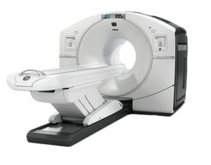
GE Discovery MI PET-CT (located in HDB)
Show More
-
The GE Discovery MI PET-CT scanner within the bic HDB facility enables high-resolution, bleeding edge research and clinical imaging capabilities. The PET detector material is a lutetium-based scintillator comprised of 19,584 total crystals apportioned into 544 blocks in 36 rings. Each crystal measures 3.95 × 5.3 × 25 mm3. The detectors themselves are silicon photomultipliers (SiPM) affixed to application-specific integrated circuits (ASICs), which provide fully digital acquisitions at much higher sensitivities than traditional analog photomultiplier tubes (PMTs). With a NEMA sensitivity of 13.7cps/kBq and a timing resolution of 385ps that enables ultra-low noise reconstructions with time-of-flight techniques, the Discovery MI achieves previously unattainable image quality. Its axial resolution is 4.8mm at 1cm from FOV center, and its transaxial resolution is 4.5mm at 10cm. The full suite of proprietary GE PET quantitation and motion artifact reduction technology is available on the scanner, including Q.Clear, Q.Freeze, and Q.Static. The CT provides a 50cm diagnostic FOV which can expand to 70cm for attenuation correction. A nominal helical pitch of 0.516 to 1.531 and a cardiac pitch of 0.16 to 0.35, allow for large axial coverage with minimal gantry rotations at low dose. The maximum X-ray power is 72kW with tube current ranging from 10 to 600mA in 5mA increments. Available scan modes include standard axial, helical, and cine techniques. Iterative image reconstructions obtain the highest resolution with the lowest possible X-ray dose.
Preclinical Imaging Scanners
-
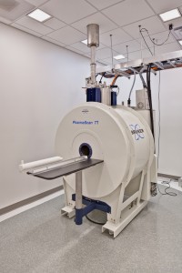
Bruker BioSpec 7.0T MRI (located in HDB)
Show More
-
The MRI is a Bruker BioSpec 7.0 Tesla, 16cm bore (B-C 70/16) platform. It is equipped with a Bruker B-GA 9S high duty cycle gradient coil, capable of delivering 30 G/cm maximum amplitude with an 80- microsecond rise (3,375 T×m-1×s-1) time along any individual axis and a up to 69 G/cm amplitude combined. The 12 shim gradient coils are integrated into the liner gradient coil, providing a total of 15 shim channels. This gradient coil provides a 9cm operational inner diameter. The system is equipped with three 1H CP volume coils and a volume transmit, surface receive RF coil set – all made by Bruker. The inner diameters of the three volume coils are 72mm, 38mm, and 23mm. The transmit-only volume resonator has an 80mm inner diameter, and the surface coil is 30mm in diameter, and both are actively-detuned. The system has two independent, fully broadband 1,000W (pulsed), 50W (CW) RF amplifiers. The receive-chain has a single broadband receiver equipped with 1H, 19F, and X filters. The system is capable of operating with either of two host computers – one running ParaVision 5.1 and the other with ParaVision 6.0. Both software configurations include all options available. The system is maintained under a parts service contract from Bruker.
-
Xenogen IVIS Spectrum (located in HDB)
Show More
-
The facility has an IVIS Spectrum system made by Xenogen/Perkin Elmer. It is capable of simultaneously imaging 5 mice under isoflurane anesthesia on a bed maintained at physiological temperature. The system has 8 excitation filters (410-760nm) and 12 emission filters (475-850nm). It is operated by a host computer running Living Image 4.1. The anesthesia is metered by an XGI-8 isoflurane vaporizer/evacuator.
-
Siemens Inveon PET-CT (located in NHB)
Show More
-
The PET-CT is a Siemens Inveon scanner. It is equipped with all available options including a variable focus X-ray source and an extra-large 165mm detector. The PET is capable of achieving 1.6 × 1.6mm pixel spacing with its 4 rings of 16 blocks (each with 20 × 20 LSO crystal arrays) and iterative reconstruction techniques. The PET detectors provide 10-bit dynamic range at 100MHz sampling and 312ps time bins.
The maximum single-position axial PET field of view is 12.7cm, but with continuous table motion, 30cm is possible. The X-ray source operates between 35-50kVp and is specified to provide a 50-micrometer focal spot. However, this particular instrument is capable of achieving ~7 micrometers isotropic CT voxels. Using a separate physiological monitoring system, it is capable of gating acquisition to the respiratory or cardiac cycle. CT-based attenuation correction is available. The host computers have all available software options and run Acquisition Workplace 4.0. There is a standalone computer that runs Research Workplace version 4.0 that can be used to process data offline.